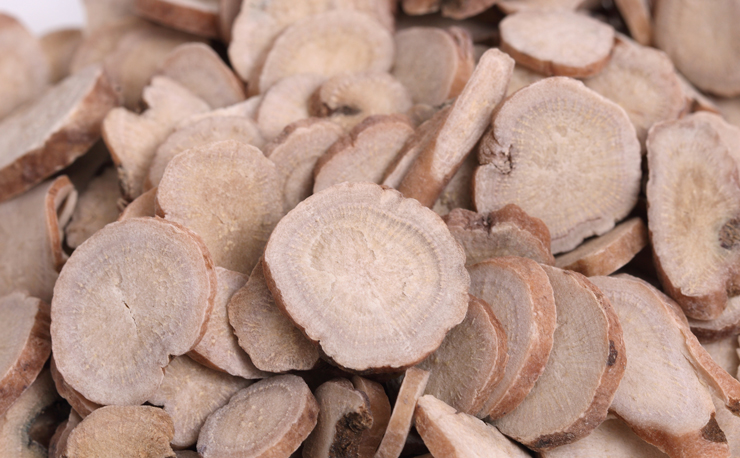
Paeoniflorin, a white peony root component, has shown inhibition of in vitro testosterone synthesis without influencing estradiol synthesis. This decreases the ovaries ‘ production of testosterone, but not the adrenal glands. Oral administration of a mixture of white peony and licorice in an ovariectomized experiment raised the accumulation of DHEA and serum estrogen. It functions as a soothing uterine smooth muscle in several species of animals. TJ-68, a traditional Japanese herbal remedy containing Paeonia lactiflora and licorice, showed effectiveness in treating amenorrhea and hyperprolactinemia caused by risperidone in one case study.
Besides, a correlation model was developed between the marker metabolites and their chemical composition to discover and test the constituents or metabolites ingested through blood postoral administration of TCM formulae. Six active components (albiflorine, paeoniflorin, coptisine, berberine, palmatine, and ginsenoside Rg1) were evaluated and screened using WXF correlation plotting between marker metabolites and serum components (PCMS).
The behavior of these compounds is currently being constantly confirmed by cell-level biological experiments. The works could provide key information for the detection, elucidation, and interpretation of the metabolism and pharmacological behavior of WXF. For the rapid discovery of potentially bioactive components and WXF metabolites, the present study was successfully applied.
The plant extract “total peony glucosides” (TGP) is a mixture of glycosides isolated from the roots of the well-known herb Paeonia lactiflora Pall. The most abundant portion and significant biologically active element of TGP is paeoniflorin (Pae). In some animal models, Pae shows strong anti-inflammatory and immune regulation activity
This study discusses Pae’s pharmacological activities and Paeoniflorin-6′-O-benzenesulfonate (CP-25) innovative anti-inflammatory and immunomodulatory agents in multiple chronic inflammatory and autoimmune disorders. Pae and CP-25’s therapeutic effects make them attractive targets for other related diseases, which in the future would require extensive work.
The TGP glycoside combination is an active plant extract extracted from the roots of traditional Chinese medicine (TCM) called Paeonia lactiflora Pall. In 1998, TGP was approved for anti-inflammatory and immunomodulatory therapy in China by the National Medical Products Administration (NMPA). It was used in the treatment of RA and Systemic Lupus Erythematosus (SLE) (Chen et al., 2013; Wang et al., 2014). In the meantime, several additional research has shown that TGP also has therapeutic value in persistent nephritis care
To improve Pae’s bioavailability, comprehensive studies have been conducted. The absorption of Pae can be significantly increased by two well-known P-glycoprotein (P-gp) antagonists, verapamil, and quinidine (Chan et al., 2006; Chen Y. et al., 2012). Modification of acetylation also increases the absorption and lipophilicity of Pae in vitro (Yang et al., 2013). It has improved and evaluated the bioavailability of benzoyl paeoniflorin sulfonate in a mouse model (Cheng et al., 2010). We prepared paeoniflorin-6′-O-benzene sulfonate (CP-25) based on this theory.
Adjuvant rat arthritis (AA) study studied Pae’s anti-inflammatory and immunoregulatory effects on mesenteric lymph node (MLN) lymphocytes and the underlying mechanisms. Pae significantly reduced arthritis scores and swelling of the secondary hind paw, pro-inflammatory cytokine output, and MLN lymphocyte proliferation. Pae mediatedβ2-adrenergic receptor expression (ADRB2) and decreased β-arrestin1/2 expression in MLN lymphocytes.
Furthermore, Pae in vitro removed MLN lymphocytes’ pro-inflammatory cAMP. For arthritis pathogenesis, Pae’s impact on desensitization of ADRB2 and β2-cAMP signal transduction in MLN lymphocytes is necessary (Wu et al., 2007). Pae also repressed malondialdehyde and caused superoxide dismutase, catalase, and glutathione peroxidase in the blood in another paper using the AA rat model. In turn, Pae blocked p65-unit, tumor necrosis factor (TNF)-α, interleukin (IL)-1β, and IL-6, and reduced COX-2 (Jia and He, 2016). Such reports suggested Pae’s reduction in AA rat illness.
The inhibition levels for the development of NO, PGE2, TNF-α andIL-6 were 17.61, 27.56, 20.57, and 29.01 percent for paeoniflorin and 17.35, 12.94, 15.29, and 10.78 percent for albiflorin relative to the LPS-induced community. Paeoniflorin and albiflorin IC50 values for NO output were 2.2 −10−4 mol / L, respectively, and 1.3 −10−2 mol / L. COX-2 protein expression was decreased with paeoniflorin by 50.98 percent and with albiflorin by 17.21 percent. The gene expression activation levels for iNOS, COX-2, IL-6, and TNF-α were 35.65, 38.08, 19.72, and 45.19 percent for paeoniflorin, respectively, and 58.36, 47.64, 50.70, and 12.43 percent for albiflorin.


